Microarray during pregnancy
Use of DNA microarray as a diagnostic tool during pregnancy: one step ahead in chromosome analysis

How can we help you?
Non-obligation guidance
Prenatal genetic diagnosis using microarray makes it possible to identify a future child’s whole genome. It displays small increases or decreases in genetic material that a karyotype does not detect. Severe genetic syndromes in foetuses that can compromise quality of life can also be detected.
What does it entail?
Prenatal genetic diagnosis using microarray makes it possible to identify a future child’s whole genome. It displays small increases or decreases in genetic material that a karyotype does not detect. Severe genetic syndromes in foetuses that can compromise quality of life can also be detected.
Progress in molecular cytogenetics has facilitated development of a ground-breaking technique: DNA microarray (aCGH). This diagnostic technique is much more sensitive and efficient than a conventional karyotype. It reaches levels of diagnosis that are 10 times superior to a karyotype. As such, it allows us to detect small changes in genetic material that cannot be detected using conventional karyotype techniques. DNA arrays for prenatal studies can also include an analysis of certain genetic syndromes that can affect a baby’s quality of life or explain previously unexplained malformations or abnormalities that were detected during pregnancy.
What is it used for? What is its purpose?
Each species has a specific chromosomal make-up. Excesses or deficiencies in that chromosomal make-up lead to abnormalities that can affect a person’s health to a greater or lesser degree depending on which region is abnormal.
DNA microarray (aCGH) allows us to understand if a foetus has all the genetic information that is found in human beings or if he or she has an excess or defect of any kind that might explain developmental malformations or abnormalities that were detected during pregnancy.
What does it entail?
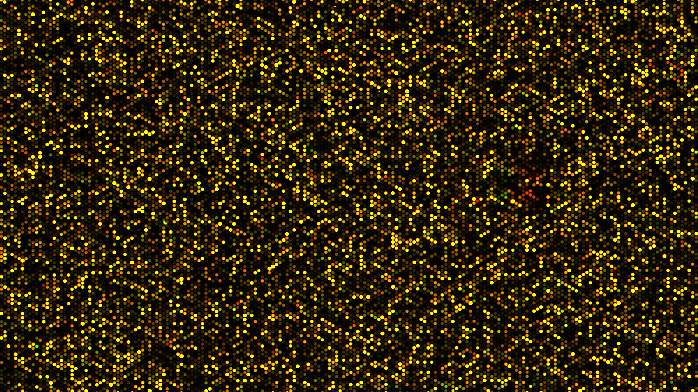
Amniocentesis or chorionic villus sampling needs to be performed in order to obtain samples of the foetus. They are used to extract DNA in order to perform the technique that consists of collective hybridisation green fluorescent colouring of the future child’s DNA. At the same time, a control DNA (with no chromosomal abnormalities) is also marked in red. Hybridisation of the mix of both fluorescent markers is performed against a DNA ‘chip’. The chip or array contains a collection of DNA molecules that sweep the entire human genome. The hybridisation result is analysed using a scanner. When DNA content is normal, the scanner detects a yellow colour (a mix of red and green). On the other hand, if there is an excess or deficiency in a chromosome region, the scanner detects the red or green colour, respectively.
When is a DNA microarray analysis advisable?
An array CGH is recommended when an abnormality such as a heart condition has been detected in a foetus using ultrasound scan markers. The analysis can also be performed when a patient is due to undergo invasive diagnosis. In cases of intrauterine foetal death or foetal loss, supplementing the cytogenetic analysis with an array CGH is also advised.
What happens if there is an abnormality in the microarray?
Appropriate genetic guidance by qualified personnel is advisable so that the findings can be explained. The couple’s options vary depending on the results that are obtained and the abnormalities found in the ultrasound scans performed on the foetus. These include both conservative and invasive options. It is sometimes necessary so analyse the parents in order to find out if the abnormality has been inherited and, as such, to understand how badly affected the individual is.